
Technology
Transformative tech, radiating from one magnet.
A new magnet architecture moves the complexity of MRI out of expensive hardware and into signal processing and reconstruction. Everything else builds on that.
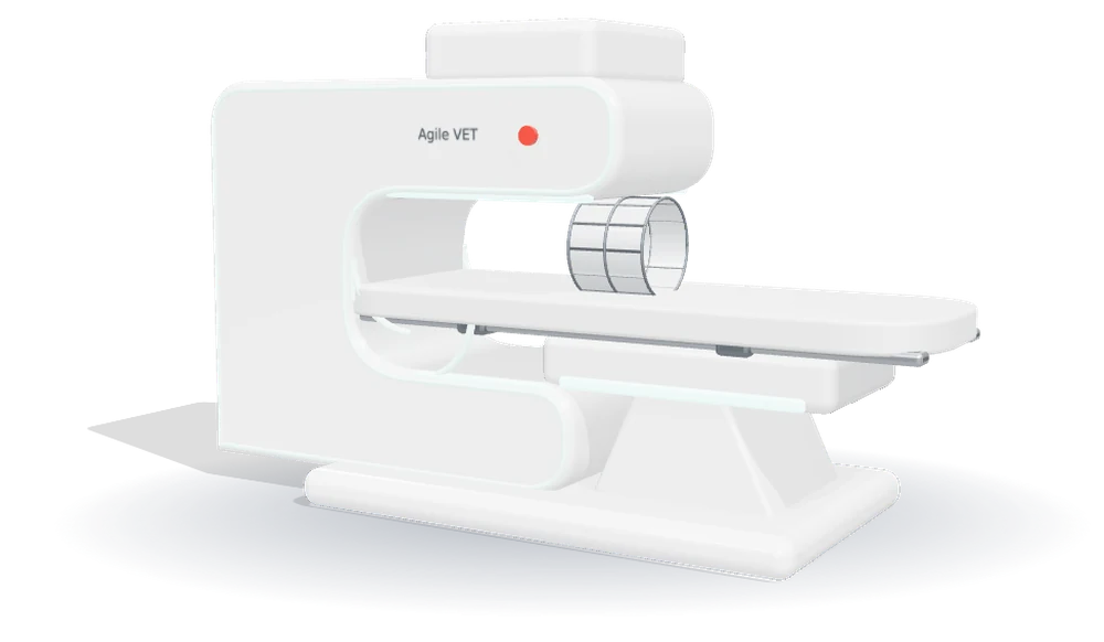 Design concept render
Four pillars, one architecture.
Smart Magnet
RF Efficiency
Robust Imaging
AI-Assisted Workflow
Why portable MRI stayed impossible
A conventional MRI scanner is expensive for reasons that compound. A strong, exquisitely uniform field needs a superconducting magnet; a superconducting magnet needs liquid helium and a cryogen service contract; the fringe field needs a shielded room; the gradient coils need power and cooling; and the whole assembly needs a dedicated, structurally reinforced space and a trained operator. Each requirement is reasonable on its own. Together they put a floor under the cost of the machine that has barely moved in forty years.
That floor is why veterinary MRI is concentrated in a handful of hospitals, why a study commonly costs a client several thousand dollars, and why so many patients are treated empirically instead.
The change
While researching MRI at Tel Aviv University, Professor Uri Nevo asked whether clear images could be produced without expensive gradient coils or a perfectly uniform field. Signal-processing work by David Corcos turned that question into acquisition methods and reconstruction algorithms, and demonstrated that meaningful MR images can be formed even in a highly distorted field.
The consequence is that complexity moves out of hardware and into software. A magnet that does not have to be perfect does not have to be superconducting, does not need helium, and does not need a room built around it.
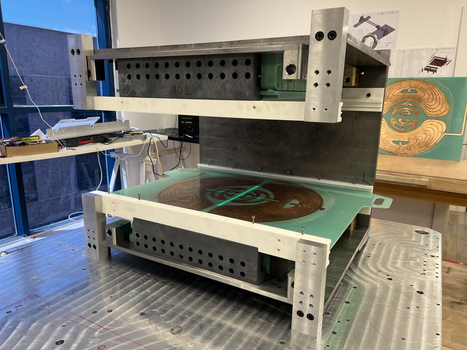
Not just renders
The magnet is real, and on the bench.
The magnet platform is being built and tested in the Agile lab today — the same architecture that will carry the veterinary configuration. Product imagery on this site is concept rendering; this photograph is the hardware behind it.
What low field can and cannot do
Low-field MRI is not high-field MRI. Signal-to-noise ratio scales with field strength, and no amount of reconstruction removes that fact. A 0.05T system will not resolve what a 1.5T system resolves, and we do not claim it will. Anyone telling you otherwise is selling something.
What has changed is where the useful threshold sits. Reconstruction and acquisition methods have moved far enough that a low-field study can answer a well-posed clinical question — is there a mass, is there a disc extrusion, is this tendon torn — in cases where the practical alternative today is no imaging at all.
Where we expect it to be useful
- Screening and triage, to decide which patients justify a high-field study
- Large, well-contrasted soft-tissue findings in a defined region — effusions, middle- and inner-ear fluid, tendon and ligament injury
- Serial follow-up, where the comparison matters more than absolute resolution
- Cases where the realistic alternative is empirical treatment without imaging
Where it will not be
- Fine structural detail at the limit of high-field resolution
- Studies where a subtle negative finding must be excluded with confidence
- Anything requiring contrast protocols and sequences not yet validated on this platform
- Any clinical decision, today, on a live patient — the veterinary configuration is not a cleared device
What is still open
The veterinary configuration is in design. Species range, patient size limits, coil geometry, the anatomical regions covered and the anaesthesia protocol are all decisions that have not been finalised, and they are precisely the decisions that veterinary clinicians should be making with us rather than being handed afterwards.
If you have a view on any of it — especially a sceptical one — we would like to hear it.
Want to see it work?
Leave your address and you will be first to see the veterinary system in action — and first to be offered a unit to test. Questions about the physics, the protocols or the field strength are welcome; a clinician here will answer them personally.
First units expected to be available to test towards the end of 2027. A few emails a year, nothing else, unsubscribe any time.